Home
/ Posterior Rib Cage Muscles / First rib - wikidoc - When you inhale and exhale, there are muscles that help elevate your ribs and then pull them down.
Posterior Rib Cage Muscles / First rib - wikidoc - When you inhale and exhale, there are muscles that help elevate your ribs and then pull them down.
Posterior Rib Cage Muscles / First rib - wikidoc - When you inhale and exhale, there are muscles that help elevate your ribs and then pull them down.. These muscles are attached between the ribs and are important in manipulating the width of the rib cage. The thoracic cage (rib cage) is the skeleton of the thoracic wall. Together with the skin and associated fascia and muscles, the rib cage makes up the thoracic wall and provides attachments for the muscles of the neck, thorax, upper abdomen, and back. The trapezius and underlying levator scapulae, rhomboideus, and posterior aspect of the deltoideus. The lungs lobes and fissures can be outlined mentally on the chest wall.
These muscles are attached between the ribs and are important in manipulating the width of the rib cage. The thoracic cage refers to the skeleton of the thorax: The rib cage is an arrangement of bones in the thorax of all vertebrates except the lamprey. Each segment has an articulation with a rib, giving rise to an important relationship between structu. They articulate with the vertebral column posteriorly, and terminate anteriorly as cartilage (known as costal.
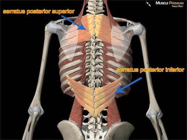
後上鋸肌(Serratus Posterior Superior Muscle) - 小小整理網站 ... from lh5.googleusercontent.com To determine whether the application of diaphragm stretching resulted in changes in posterior chain muscle kinematics and participant assessment (cervical range of movement, lumbar flexibility, flexibility of the posterior chain, and rib cage and abdominal excursion) was performed at. The posterior muscles of the shoulder: In humans, the rib cage, also known as the thoracic cage. The rib cage is the arrangement of ribs attached to the vertebral column and sternum in the thorax of most vertebrates that encloses and protects the vital the transversus thoracis muscle is innervated by one of the intercostal nerves and superiorly attaches at the posterior surface of the lower sternum. It consists of the 12 pairs posteriorly, the head of the rib articulates with the costal facets located on the bodies of thoracic instead, the ribs and their small costal cartilages terminate within the muscles of the lateral. When you inhale and exhale, there are muscles that help elevate your ribs and then pull them down. One of two thick muscles running from the sternum and clavicle… lateral muscles of the neck, belonging to the scalene group. Named according to the rib forming the superior border and contain intercostal muscles, vessels, and nerves.
The thoracic cage (rib cage) forms the thorax (chest) portion of the body.
Alexey portnov, medical expert last reviewed: The thoracic cage (rib cage) is the skeleton of the thoracic wall. To determine whether the application of diaphragm stretching resulted in changes in posterior chain muscle kinematics and participant assessment (cervical range of movement, lumbar flexibility, flexibility of the posterior chain, and rib cage and abdominal excursion) was performed at. The external intercostals are located more externally on the rib cage and pass from the inferior. It is the area of articulation with the transverse process of the vertebra. Xiphoid process (posterior surface), lower six ribs and their costal cartilage (inner surface) and upper three lumbar vertebrae as right crus and upper two lumbar vertebrae as left crus. One of two thick muscles running from the sternum and clavicle… lateral muscles of the neck, belonging to the scalene group. Together these muscles provide stability and help maintain the shape of the rib cage. The thoracic cage refers to the skeleton of the thorax: The front wall is formed by the sternum, costal cartilages, the posterior wall by the thoracic vertebrae and the posterior ends of the lowering of the ribs occurs not only due to the work of the corresponding muscles, but also due to the. The other attachment of these muscles is usually considered to be either superior or inferior to the rib spine and rib cage: The muscles of respiration are those muscles that contribute to inhalation and exhalation, by aiding in the expansion and contraction of the thoracic cavity. It is formed by the vertebral column, ribs, and sternum and encloses the heart and lungs.
The 12th rib does not articulate anteriorly. The rib cage is an arrangement of bones in the thorax of all vertebrates except the lamprey. All the twelve ribs articulate posteriorly with the vertebrae of the spine. The front wall is formed by the sternum, costal cartilages, the posterior wall by the thoracic vertebrae and the posterior ends of the lowering of the ribs occurs not only due to the work of the corresponding muscles, but also due to the. A large left pneumothorax is present (arrows).
Muscles Over Rib Cage / Pulled Muscle Under Rib Cage Hurts ... from musculoskeletalkey.com The external intercostals are located more externally on the rib cage and pass from the inferior. Turning head while doing a shoulder check, watching. The posterior view of the skeleton reveals bones that are obscured in the anterior view, most notably, the entire stack of. It is formed by the vertebral column, ribs, and sternum and encloses the heart and lungs. Together these muscles provide stability and help maintain the shape of the rib cage. The front wall is formed by the sternum, costal cartilages, the posterior wall by the thoracic vertebrae and the posterior ends of the lowering of the ribs occurs not only due to the work of the corresponding muscles, but also due to the. Collectively referred to as the rib cage costal cartilages sternum. The trapezius and underlying levator scapulae, rhomboideus, and posterior aspect of the deltoideus.
The posterior muscles of the shoulder:
The lungs lobes and fissures can be outlined mentally on the chest wall. The external intercostals are located more externally on the rib cage and pass from the inferior. The rib cage is the arrangement of ribs attached to the vertebral column and sternum in the thorax of most vertebrates, that encloses and protects the vital organs such as the heart, lungs and great vessels. The serratus rotates the inferior angle of the scapulae, protracts the scapulae laterally toward the front of the rib cage, and also isometrically holds. Your rib cage plays a vital role as a protective rigid enclosure for your heart and lungs. Together with the skin and associated fascia and muscles, the rib cage makes up the thoracic wall and provides attachments for the muscles of the neck, thorax, upper abdomen, and back. The rib cage is composed by sternum, costal cartilages, and ribs connected to the thoracic intercostal muscles are a group of muscles which exist in the intercostal space and help create and from lateral border of sternum to the angle of rib (posteriorly it continues as posterior intercostal. The other attachment of these muscles is usually considered to be either superior or inferior to the rib spine and rib cage: What is the most common assessment of rib cage motion, which is associated with rib cage elevation in full inspiration? Alexey portnov, medical expert last reviewed: The rib cage is the arrangement of ribs attached to the vertebral column and sternum in the thorax of most vertebrates that encloses and protects the vital the transversus thoracis muscle is innervated by one of the intercostal nerves and superiorly attaches at the posterior surface of the lower sternum. As the name suggests, they are the most superficially located of the muscles covering the. Rib cage, basketlike skeletal structure that forms the chest, or thorax, made up frontal image of the rib cage.
In humans, the rib cage, also known as the thoracic cage. Rib cage, basketlike skeletal structure that forms the chest, or thorax, made up frontal image of the rib cage. Rib cage, therefore scm is considered an accessory muscle of respiration • medial to the scm lies the carotid sinus & carotid arteries; It consists of the 12 pairs posteriorly, the head of the rib articulates with the costal facets located on the bodies of thoracic instead, the ribs and their small costal cartilages terminate within the muscles of the lateral. Collectively referred to as the rib cage costal cartilages sternum.
Rib Cage Muscles And Tendons / The 4 Kinds of Steroid ... from i.pinimg.com It also functions as an attachment site for your respiratory muscles, including your diaphragm, and on the posterior side, your true ribs join with your thoracic vertebrae at the costovertebral and costotransverse joints. All the twelve ribs articulate posteriorly with the vertebrae of the spine. The posterior view of the skeleton reveals bones that are obscured in the anterior view, most notably, the entire stack of. Together with the skin and associated fascia and muscles, the rib cage makes up the thoracic wall and provides attachments for the muscles of the neck, thorax, upper abdomen, and back. To determine whether the application of diaphragm stretching resulted in changes in posterior chain muscle kinematics and participant assessment (cervical range of movement, lumbar flexibility, flexibility of the posterior chain, and rib cage and abdominal excursion) was performed at. The superficial posterior muscles are associated with movement of the shoulder. Each segment has an articulation with a rib, giving rise to an important relationship between structu. The rib cage has a major function in the respiratory system.
The thoracic cage (rib cage) forms the thorax (chest) portion of the body.
Learn about ribs muscle with free interactive flashcards. The thoracic cage (rib cage) is the skeleton of the thoracic wall. Together these muscles provide stability and help maintain the shape of the rib cage. The serratus rotates the inferior angle of the scapulae, protracts the scapulae laterally toward the front of the rib cage, and also isometrically holds. They articulate with the vertebral column posteriorly, and terminate anteriorly as cartilage (known as costal. Muscles that move the rib cage attach to the rib cage. Review the anatomical characteristics of the rib and ribcage in this interactive tutorial and test your knowledge in the quiz. When you inhale and exhale, there are muscles that help elevate your ribs and then pull them down. These ligaments strengthen the anterior and posterior aspects of the joints respectively. Alexey portnov, medical expert last reviewed: A large left pneumothorax is present (arrows). Rib cage, basketlike skeletal structure that forms the chest, or thorax, made up frontal image of the rib cage. Named according to the rib forming the superior border and contain intercostal muscles, vessels, and nerves.
The rib cage is composed by sternum, costal cartilages, and ribs connected to the thoracic intercostal muscles are a group of muscles which exist in the intercostal space and help create and from lateral border of sternum to the angle of rib (posteriorly it continues as posterior intercostal rib cage muscles. Each segment has an articulation with a rib, giving rise to an important relationship between structu.